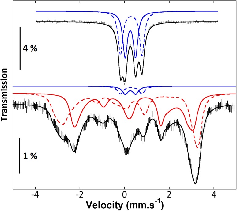
Fig. 1.
Characterization by Mössbauer spectroscopy of oxidized (top) and dithionite-reduced (bottom) human mitoNEET recorded at 4.2 K in a magnetic field of 600 G applied parallel to the direction of the γ-rays. The solid and dashed blue lines represent the contributions of [2Fe-2S]2+ clusters, and the solid and dashed red lines represent the contributions of [2Fe-2S]+ clusters. From [15]

