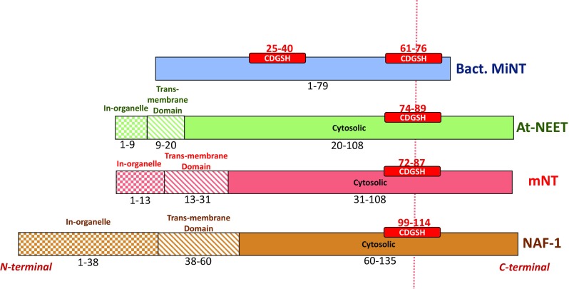
Fig. 1.

NEET proteins CDGSH organization. The location of the CDGSH domain(s) is shown in (red box) bacterial MiNT (blue), At-NEET (green), mitoNEET (red) and NAF-1 (brown). Different textures of the boxes were used to distinguish between different domains: in-organelle domain (checker texture), inter-membrane domain (diagonal lines pattern) and cytosolic domain (full color). The sequence interval is reported for each domain. The different regions specified here are based on the sequence of each protein
