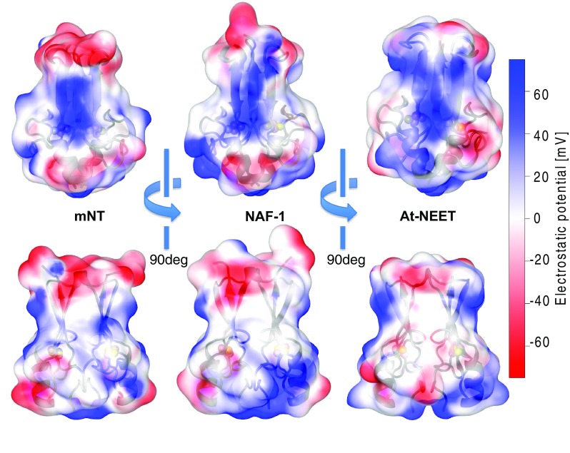
Fig. 4.

Electrostatic potential on the NEET protein’s surface. The electrostatic potential values (estimated using NEET proteins’ force files [85] and APBS electrostatic [103]) of mNT [25], NAF-1 [38] and At-NEET [30] are here reported over each protein surface. The side facing the plane of the β-sheet (top) and the side view (bottom) are here reported. The color code refers to the electrostatic potential values reported on the right
