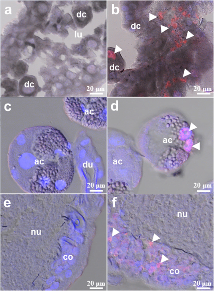
Figure 3.
Localization of the Thogoto virus (THOV) in the midgut (a,b), salivary glands (c,d), and synganglia (e,f) of partially fed, infected adult ticks via anal pore microinjection. Viral antigens were detected using a specific THOV polyclonal antibody (b,d,f), while normal mouse serum served as a control (a,c,e). Nuclei counterstaining (blue) was done using DAPI, and arrowheads denote THOV antigens (red) (bar = 20 μm). dc: digestive cells; ac: acinus; du: duct; lu: lumen; nu: neuropile; co: cortex.

