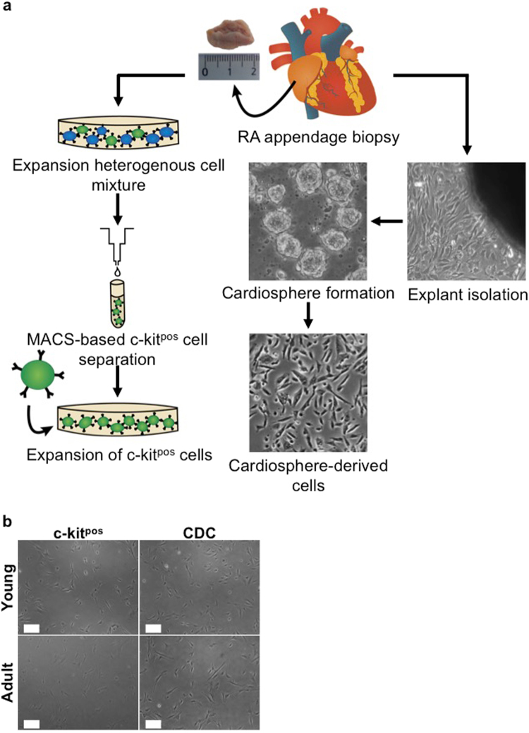
Figure 1.
Isolation and morphological comparison of c-kitpos CPCs and CDCs. (a) Schematic overview of the steps involved to isolate c-kitpos CPCs and CDCs. To isolate c-kitpos cells, a human RA appendage biopsy is minced into small pieces and collagenase digested to culture a heterogeneous mixture of single cells for 2 passages prior to MACS-based selection. To obtain CDCs, a mild collagenase-based digestion is performed on small myocardial fragments to allow outgrowth of fibroblasts and phase-bright cells from the cardiac explants. The latter are transferred onto a poly-D-Lysine coated dish to form cardiospheres. Finally, the cardiospheres are collected and grown as monolayer cells called CDCs. (b) Phase bright-field pictures of c-kitpos CPCs and CDCs derived from young and adult donors. Scale bars represent 200 µm. CPC, cardiac progenitor cell; CDC, cardiosphere-derived cell; RA, right atrial.

