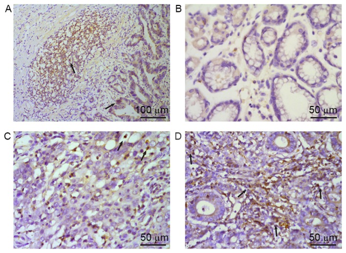
Figure 4.

Expression of IL-22 protein in tissue specimens from patients with gastric cancer was evaluated using immunohistochemistry. IL-22-positive cells presented brown-yellow staining in the cytoplasm, as indicated by arrows. (A) IL-22 was mainly distributed in tumor cells and infiltrating lymphocytes around the tumor. IL-22 expression in (B) normal gastric mucous tissue and (C) tumor tissues in Stage I–II and (D) Stage III–IV gastric cancer. Hematoxylin staining of the same paraffin sections was used to distinguish between tumor cells and infiltrating lymphocytes. IL, interleukin.
