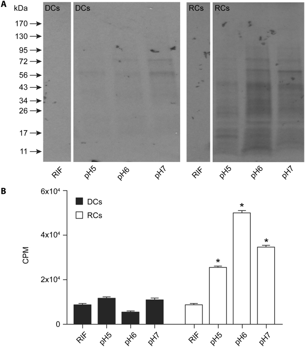
Figure 4.
Impact of pH variations assessed on the 35S Cys-Met incorporation into E. chaffeensis cell-free organisms. (A) Autoradiography imaging was performed to assess the impact of three different pH units; 5, 6 and 7 for DCs and RCs. In the first lane, we included rifampin (RIF) containing sample with the media at pH 5.0 to serve as a negative control (B). As in panel A, except that the scintillation counting data were presented. Significant change noted relative to RIF controls are identified with a *above each bar. (Note: Original images used in preparing Figure 4A were provided as the Supplementary information file.)

