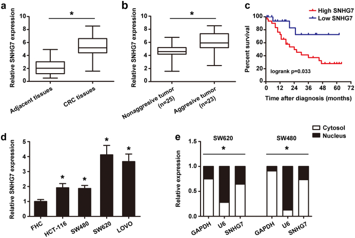
Fig. 1. The differential expression of SNHG7 in CRC tissues and cell lines and subcellular location.
a The differential expression of SNHG7 in CRC samples (n = 48) and adjacent normal colon tissues (n = 48) was shown. b The differential expression of SNHG7 in CRC patients with liver metastasis (n = 23) and without metastasis (n = 25) was analyzed. c Kaplan−Meier analyses of the correlations between SNHG7 level and overall survival of 48 patients with CRC (p = 0.033; log-rank test). The median of the dataset is selected as the cutoff point between “High SNHG7” and “Low SNHG7”. d The differential levels of SNHG7 in CRC cell lines and FHC cell were examined. e Cellular localization of SNHG7 in CRC cells was shown. GAPDH and U6 served as a cytoplasmic and nuclear localization marker, respectively. The error bars in all graphs represented SD, and each experiment was repeated three times. *p < 0.05

