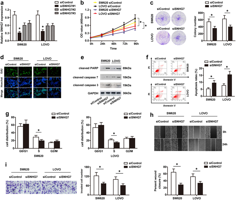
Fig. 2. Knockdown of SNHG7 inhibited cell proliferation, migration and invasion, and promoted apoptosis of CRC cells in vitro.
a The level of SNHG7 transfection with siSNHG7 or siControl was analyzed by qRT-PCR. b Growth curves of SW620 and LOVO cells after transfection with siSNHG7 or siControl were determined via CCK-8 assays. c Colony formation assays showed that knockdown of SNHG7 inhibited CRC cell proliferation. d Suppression of SNHG7 expression attenuated the proliferation of CRC cells by EdU assay. Scale bars = 50 μm. e The levels of cleaved PARP, cleaved caspase-7, and cleaved caspase-3 following SNHG7 silenced in CRC cells were determined via western blot. f Flow cytometry assay showed that silencing of SNHG7 increased the rate of apoptosis in CRC cells. g Flow cytometry assay showed that siSNHG7 resulted in S arrest in CRC cells. The cell cycle distribution was exhibited. h siSNHG7 resulted in a slower closing of scratch wound by wound-healing assay. Scale bars = 50 μm. i Transwell invasion assay was measured and the results were expressed as the number of invaded cells per field. Scale bars = 20 μm. The error bars in all graphs represented SD, and each experiment was repeated three times. *p < 0.05

