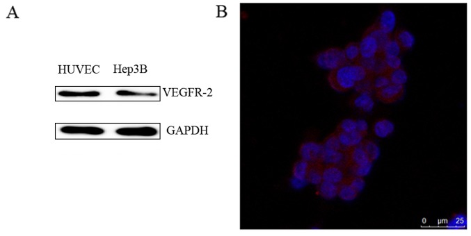
Figure 2.
The expression of VEGFR-2 in Hep3B cells. (A) Representative western blot image of VEGFR-2 expression in Hep3B cells. (B) Representative immunofluorescence image of Hep3B cells to examine the localization of VEGFR-2 (Scale bar=25 µm). VEGFR-2, vascular endothelial growth factor receptor-2. The blue fluoresence represents the nucleus and the red fluoresence represents the expression of VEGFR-2.

