. 2016 Feb 12;7(5):3279–3285. doi: 10.1039/c6sc00057f

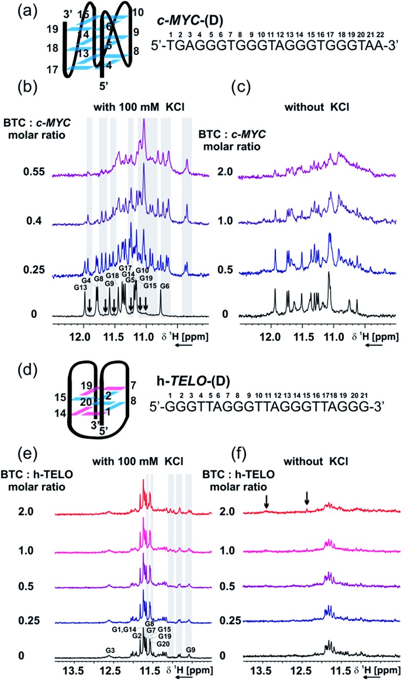
Fig. 4. NMR titration of c-MYC-(D) and h-TELO-(D) in the presence and absence of BTC and K+. (a) Scheme of the major conformation of c-MYC-(D), as determined using NMR from Ambrus et al.43 [PDB code: ; 1XAV], with sequence and numbering. Imino region of the 1D 1H NMR spectrum of c-MYC-(D) in the presence of an increasing amount of BTC, with (b) or without (c) 100 mM KCl. (d) Scheme of the proposed structure of h-TELO-(D), with sequence and numbering. Imino region of the 1D 1H NMR spectrum of h-TELO-(D) in the presence of an increasing amount of BTC, with (e) or without (f) 100 mM KCl. Experimental conditions: 100 μM DNA, 25 mM Tris–HCl buffer (pH 7.4), 298 K, 600 MHz. Guanine residues in anti and syn conformations are represented in cyan and magenta, respectively.

Fig. 4