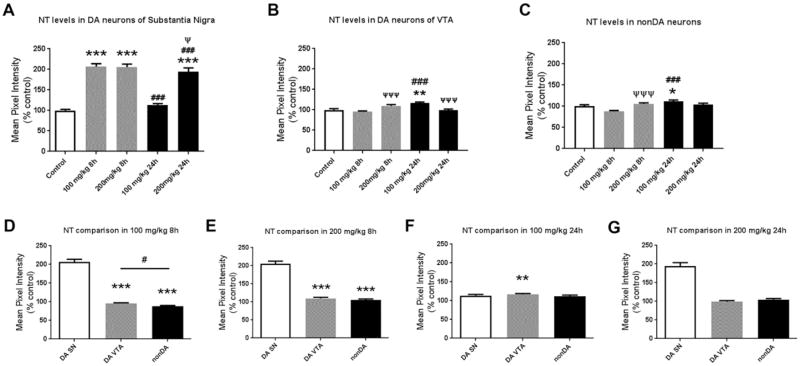
Figure 5. Quantitative analysis of cellular nitrotyrosine levels shows that PhIP treatment produces oxidative damage in dopaminergic neurons of substantia nigra relative to other cell populations.
To quantify oxidative damage, ROIs surrounding cell bodies of dopaminergic (DA) neurons (TH+) in substantia nigra (SN) were drawn and NT intensity in each ROI was quantified, and normalized to the mean of the control (A). The data are presented as the mean ± SEM (n = 390–671 cell bodies from 5 animals/treatment). NT levels were also quantified in DA neurons from the ventral tegmental area (VTA) (B) and non-dopaminergic (nonDA) neurons localized dorsally to the SN (C) (n = 162–287 bodies from 3 animals/treatment and n = 129–325 cell bodies from 3 animals/treatment, respectively). *p<0.05, **p<0.01, ***p<0.001 compared to control; ###p<0.001 compared to same-dose group; Ψp<0.05, ΨΨΨp<0.001 compared to same-time group; Kruskal-Wallis test followed by Dunn’s multiple comparisons post hoc test. To directly compare the magnitude of NT level changes for each of the three cell populations, normalized values were analyzed separately for each dose and time-point (D–G). **p<0.01, ***p<0.001 compared to DA SN; #p<0.05 compared to indicated group; Kruskal-Wallis test followed by Dunn’s multiple comparisons post hoc test.

