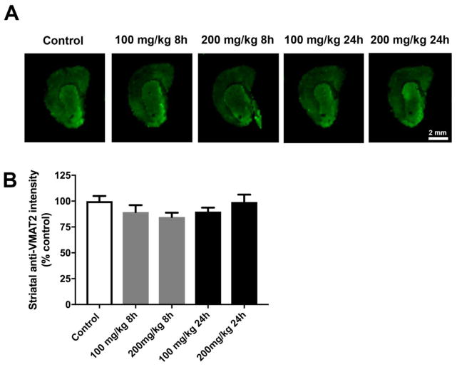
Figure 6. Acute PhIP treatment does alter striatal VMAT2 levels.
Coronal sections from the striatum from control and PhIP-treated animals were immunofluorescently stained for VMAT2 (A). Immunofluorescent intensity of ROIs surrounding the striatum was quantified using Image Studio version 3.1. All quantifications were normalized to average of control group (B). Data are presented as mean ± SEM (n = 4 or 5 sections/animal from 5 animals per treatment).

