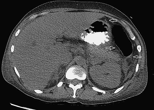
Fig. 2.

CT scan (abdomen) showing thickening of the wall of the stomach that could be related to the patient's pancreatitis. There is peripancreatic fluid (black arrow) and fluid adjacent to the splenic flexure, the inferior aspect of the spleen, and left pericolic gutter (white arrows). The pancreas itself is symmetrically enhanced with no evidence of pancreatic necrosis, peripancreatic abscess/hemorrhage, or pancreatic mass.
