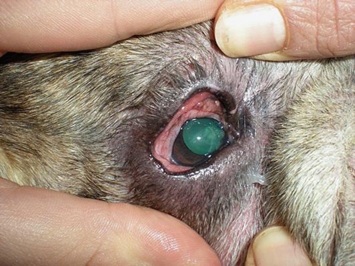
Fig. 6.

Same eye of Figure 5 after 10 days of therapy. Corneal edema has resolved and neovascularization remains only with a single, small caliper vessel arising from the limbus at the level of the medial cantus. Entropion and mucous discharge are still present.
