Fig. 1.
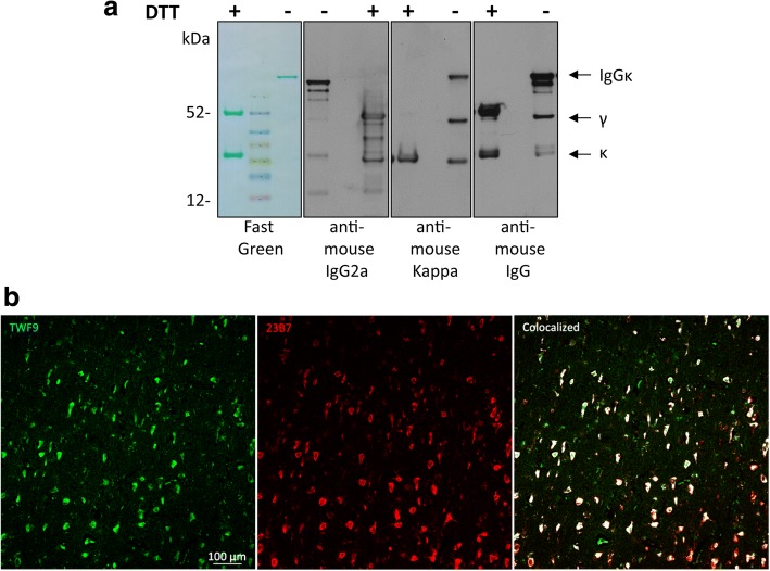
Characterization of TWF9, an anti-β-sheet conformation antibody. a TWF9 under reducing (+DTT) and nonreducing (−DTT) conditions. Left panel: fast green reversible protein stain; second panel: anti-mouse gamma 2a specific antibody; third panel: anti-mouse kappa light chain specific antibody; right panel: anti-mouse IgG antibody. b Representative image showing staining patterns between GW-23B7 [27] (red) and TWF9 (IgG) (green). Areas of colocalization are shown in white

