Fig. 7.
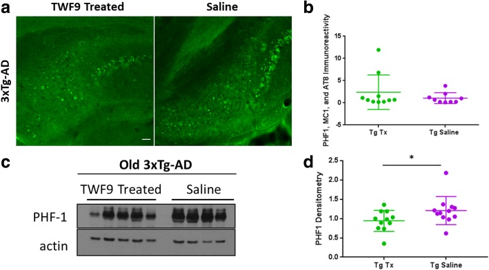
TWF9 treatment reduces soluble levels of phosphorylated tau. a Representative images of phosphorylated tau immunoreactivity in subiculum of old 3xTg-AD mice. Scale bar = 100 μm. b Analysis of tau immunoreactivity measured as percentage of subiculum area covered by phosphorylated tau. Quantification of mixed PHF1, MC1, and AT8 staining in the subiculum from two to four sections per mouse; n = 11 for transgenic (Tg) treated (Tx) and n = 12 for Tg saline. Data shown as mean ± SD. Analysis by two-tailed Mann-Whitney test. c Western blot analyses of tau pathology in old TWF9-treated and saline-treated 3xTg-AD mice. Abnormal hyperphosphorylated tau at tau sites (pSer396/pSer404) was measured by PHF1 in the soluble fraction of homogenized brains; n = 11 for Tg Tx and n = 12 for Tg saline. 15 μg of 3xTg-AD S2 fraction was loaded. d Quantification of Western blots normalized to actin is shown as mean ± SD. *p < 0.05 by two-tailed Mann-Whitney test

