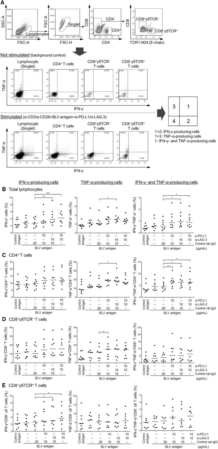
Figure 5.

Effect of PD-1/PD-L1 and LAG-3/MHC II blockade on Th1 cytokine production in BLV-specific T cells. PBMCs (n = 8; AL n = 6, PL n = 2) were cultivated with 10 µg/mL each of blocking mAbs (anti-PD-L1 and anti-LAG-3 mAbs; 10 μg/mL) or rat IgG control in the presence of FLK-BLV antigen (2%) and anti-CD3 and anti-CD28 agonist mAbs (1 µg/mL each) for 18 h. A Gating strategy and representative dot plots for expression analyses of IFN-γ and TNF-α on total lymphocytes, CD4+ T cells, CD8+γδTCR− T cells, and CD8−γδTCR+ T cells from peripheral blood of an AL animal. Values in the quadrants indicate percentages of cells. Percentages of IFN-γ+ (left), TNF-α+ (center), IFN-γ+TNF-α+ cells (right) among total lymphocytes (B), CD4+ T cells (C), CD8+γδTCR− T cells (D), and CD8−γδTCR+ T cells (E) in PBMCs treated with blocking mAbs. Bars indicate group median response. Significant differences between each group were determined using Friedman test, where P < 0.05, P < 0.01, and P < 0.001, indicated by asterisks (*, **, and ***, respectively).
