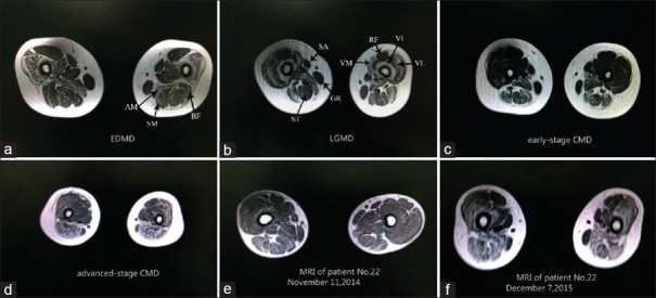
Figure 2.
Muscle MRI of patients with three different clinical phenotypes caused by LMNA gene mutations of bilateral thigh (section on one third part of thigh). (a-d) The most prominently involved muscles of these three phenotypes were the adductor magnus (AM), the semimembranosus (SM), and the long and short heads of the biceps femoris (BF). The gracilis (GR) and sartorius (SA) exhibited less infiltration. The involvement of the vasti muscles (VM), with a relative sparing of the rectus femoris (RF), is prone to be observed in EDMD, LGMD, and advanced-stage CMD; (e and f): Follow-up MRI of CMD patient. MRI: Magnetic resonance imaging; EDMD: Emery-Dreifuss muscular dystrophy; LGMD: Limb-girdle muscular dystrophy; CMD: Congenital muscular dystrophy.

