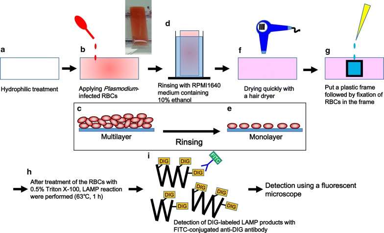
Fig. 1.
Protocol for in situ LAMP for Plasmodium-infected red blood cells on hydrophilic-treated COC plates. After hydrophilic treatment of COC plates (a), iRBCs were added to the plates, followed by 10 min standing to allow the RBCs to settle down on the plate surface (b). A picture of a plate (box). Schematic cross-sectional image of the RBCs forming a multilayer on a COC plate (c). After removing non-adherent cells (d), cells were spread as a monolayer on the plates (e). Plates were dried rapidly with a hair dryer (f). The cells were fixed with formalin (g), followed by permeabilization with Triton X-100. Then, LAMP reaction with DIG-labeled dUTP and a specific primer set for P. falciparum 18S rRNA gene was performed (h). The resulting LAMP products in the cells were visualized with FITC-conjugated anti-DIG antibody using a fluorescence microscope (i)

