Fig. 2.

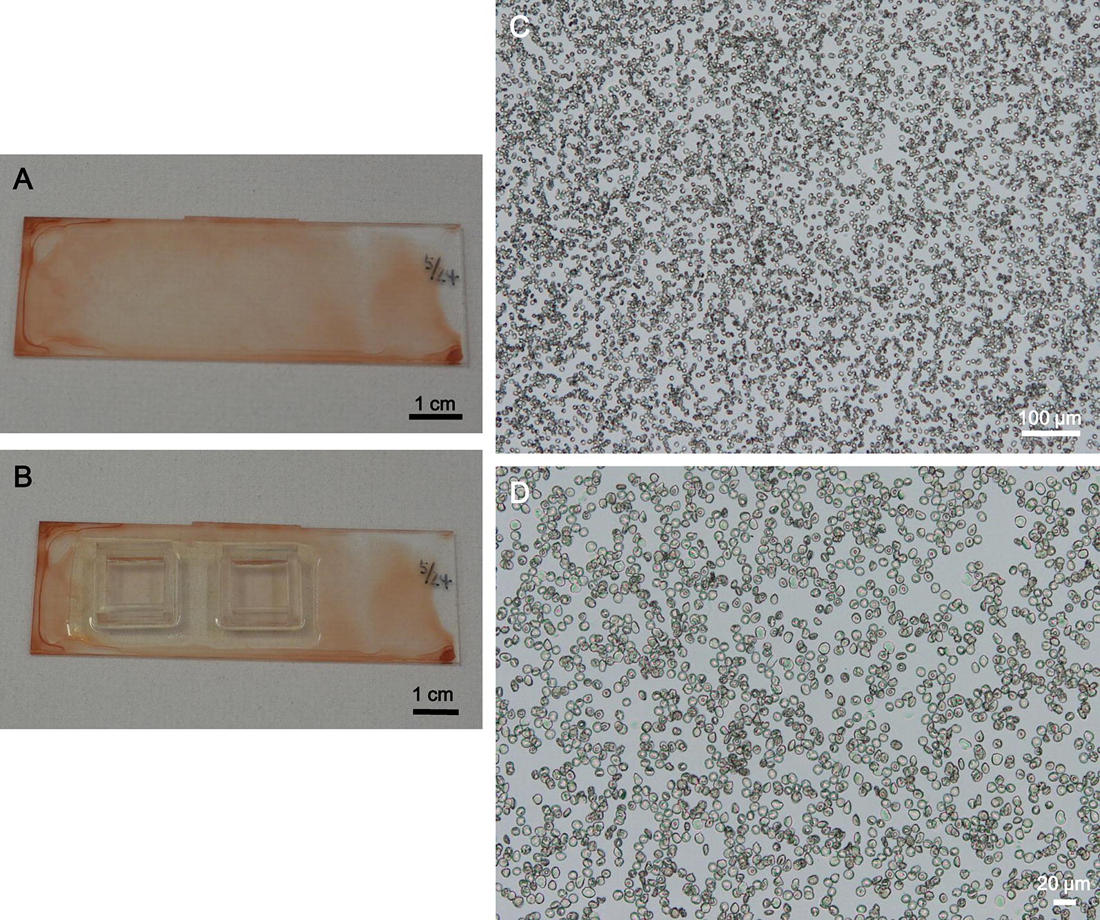
Representative images of red blood cells on hydrophilic-treated plates for in situ LAMP assay. a, b A hydrophilic-treated COC plate with dried RBCs without a plastic frame (a) and with plastic frames (b). c, d Representative microscopic images of the RBCs on the plate at ×50 magnification (c) and ×200 magnification (d)
