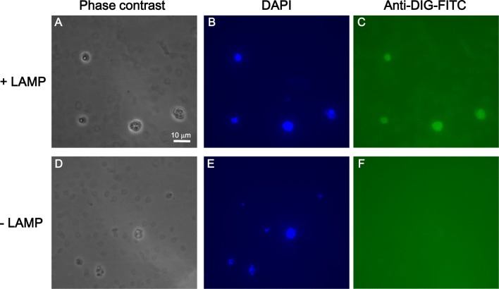
Fig. 3.
Detection of Plasmodium falciparum in red blood cells using in situ LAMP with DIG-labelled dUTP. a–c iRBCs on the hydrophilic-treated plates were analysed by in situ LAMP assay. Representative phase-contrast microscopic images (1000×, oil immersion) of the cells (a), fluorescence microscopy image in which the nucleic acid was stained with DAPI (b) and in which LAMP reaction was performed (c). d–f For a negative control, cells on the plates were handled according to the protocol described in Fig. 1, except PBS was added instead of the LAMP reaction mixture. Representative phase-contrast microscopic image of the cells (d), fluorescent microscopic image in which nucleic acid was stained with DAPI (e), and fluorescent microscopic image of cells visualized with a filter for FITC (f). Note that DAPI-positive RBCs are parasite-infected RBCs

