Fig. 4.

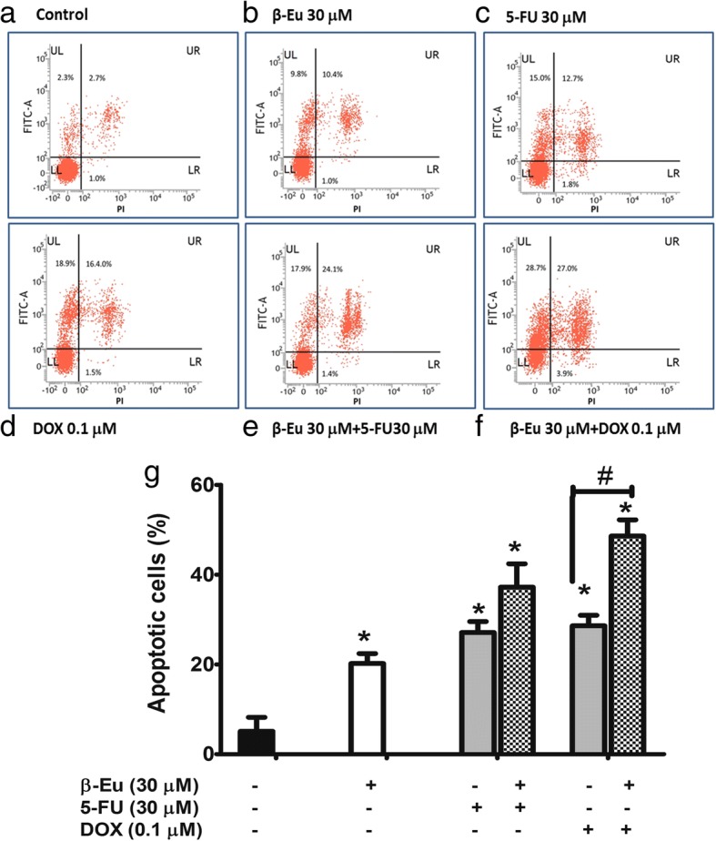
Enhancing the effect of β-eudesmol on apoptotic activities of 5-FU and DOX on CCA cells. Flow cytometry analyses of KKU-100 cells using double staining with annexin V (annexin V, vertical line) and propidium iodide (PI, horizontal line). a control, b β-eudesmol (30 μM), c 5-FU (30 μM), d DOX (0.1 μM), e combination of β-eudesmol (30 μM) with 5-FU (30 μM), and f combination of β-eudesmol (30 μM) with DOX (0.1 μM) for 24 h. Flow cytometry apoptotic results are shown in four subpopulations which indicate: early apoptotic cells (upper left), late apoptotic cells (upper right), normal cells (lower left) and necrotic cells (lower right). g The levels of KKU-100 cell apoptosis expressed as the percentage of apoptotic cells are depicted in the graph (mean ± SD averaged from three independent experiments, triplicate each). *p < 0.05 vs untreated control. #p < 0.05 vs chemotherapeutic agents alone. * indicates statistically significant difference with each drug alone (paired t-test, p < 0.05). β-Eu = β-Eudesmol, 5-FU = 5-Fluorouracil, DOX = Doxorubicin
