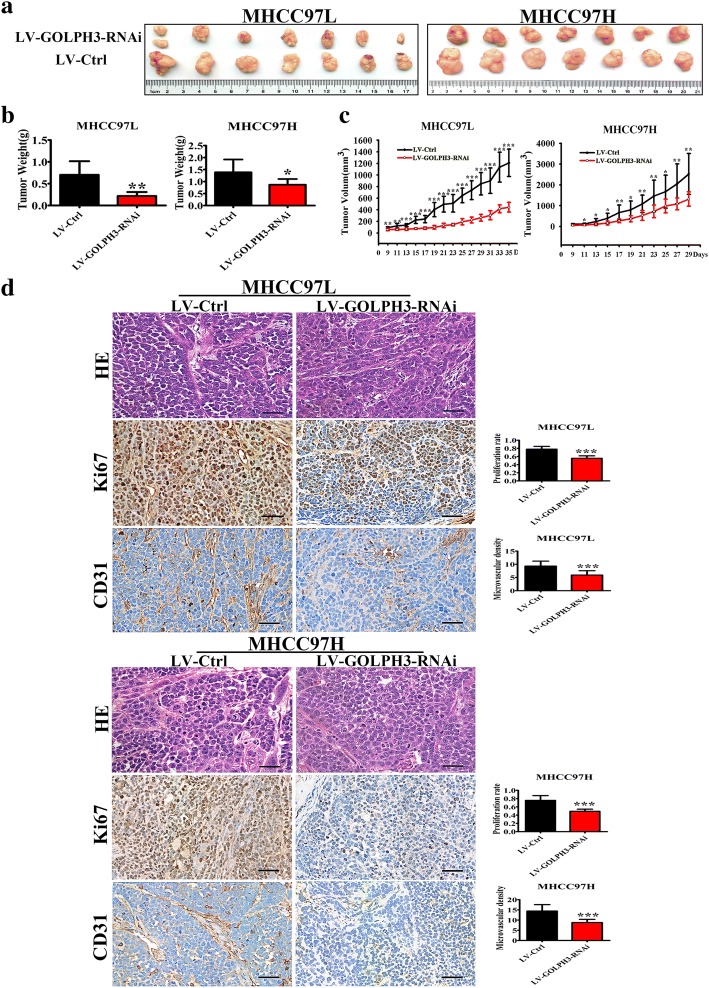
Fig. 3.
GOLPH3 contributed to the progression of HCC in vivo; a Photographs of tumors dissected from mice (n = 7) injected with LV-GOLPH3-RNAi or LV-Ctrl HCC cells. b Average tumors weight of mice from each group was shown. c Volume growth curves for the tumors formed by the HCC cells transfected with LV-GOLPH3-RNAi or LV-Ctrl. d HE staining and expression of Ki67 and CD31 in tumors tissue formed by the HCC cells transfected with LV-Ctrl or LV-GOLPH3-RNAi. The magnification of images was 400×. All experiments were performed in triplicate with at least three independent experiments. Data were presented as the mean ± SD. P-values were calculated using Student’s t-test, * P < 0.05, ** P < 0.01, *** P < 0.001, significant difference compared with the LV-Ctrl group

