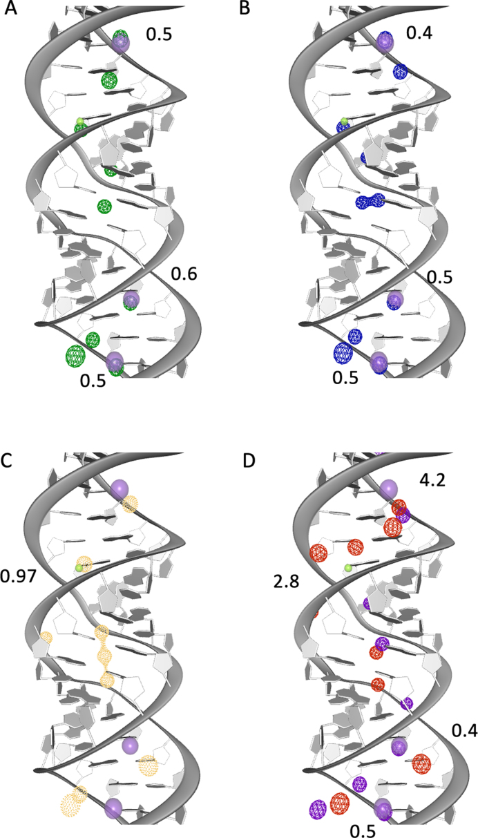
Figure 4.
Experimental and predicted ion binding sites. The helical structure (PDB id: 2QEK) has one Mg2+ (solid green) and three K+ (solid purple) binding sites. During the equilibration phase we observe Mg2+ and Na+ in close proximity to the experimentally predicted binding sites. The RMSD between experimental and predicted binding sites are given in Å. The 10 top ranked ion binding sites predicted with MobyWat for one replica for each of the four systems is shown: (A) Na+CI (green), (B) Na+PS (blue), (C) Mg2+CI (orange), (D) in Mg2+PS there are Mg2+ (red) and Na+ (purple).

