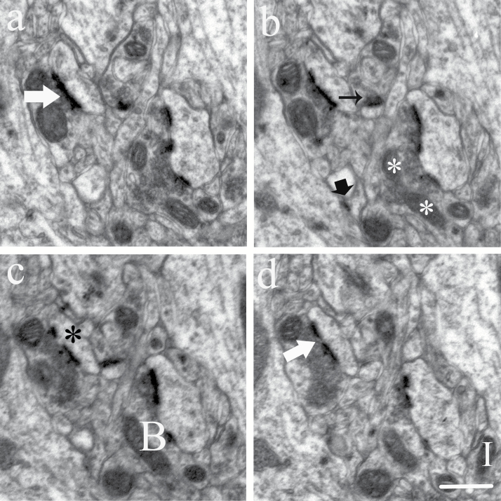
Figure 1.
Count of synapses and mitochondria in serial sections. Electron micrographs of consecutive ultrathin sections (a–d) showed nonperforated synapses (black arrow), a perforated synapse (large white arrow), and shaft synapse (large black arrow). The postsynaptic spine exhibited PSD discontinuities (black stars). Mitochondria: Isolated particle (I), A branch dividing (white stars), or branches connection (B). Scale bar, 0.5 μm.

