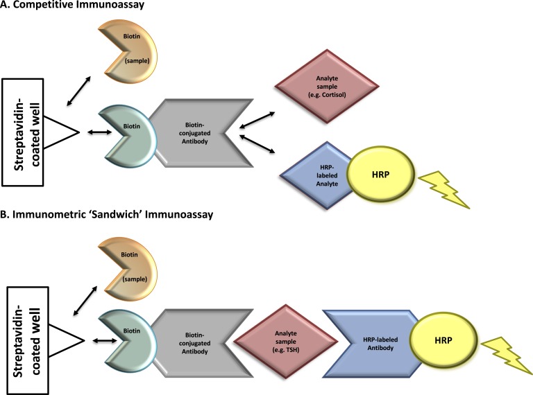
Figure 1.
Mechanism of biotin interference with immunoassay methodologies. Immunoassays in this case report contain a solid phase with a wash step that physically separates the label-bound analyte/antibody from the free label. Assays that contain this step are referred to as “heterogeneous” or “multistep” (vs homogeneous assays that do not contain a wash step). (A) Competitive immunoassays are comprised of exogenous biotin-conjugated antibodies that compete for binding with an analyte of interest in the patient’s sample as well as exogenous labeled analyte. Complexes of biotin-antibody-analyte are captured to a streptavidin-coated well through strong interactions between biotin and streptavidin. A wash step removes any unbound materials. The exogenous labeled analyte is conjugated to an enzyme [e.g., horseradish peroxidase (HRP)]. Substrate is added to the well and oxidized by horseradish peroxidase, producing a luminescence signal, measured by spectrophotometry. The measured signal is inversely proportional to the concentration of analyte in the patient’s sample. Elevated concentrations of biotin in a patient’s sample can compete with biotin-antibody-(labeled) analyte complexes for binding to the streptavidin-coated well. This leads to the detection of a diminished signal causing a falsely high analyte result. (B) Immunometric “sandwich” immunoassays contain an exogenous biotin-conjugated antibody and exogenous labeled antibody. Both antibodies bind to the same analyte of interest, forming a “sandwich”. Biotin-antibody-analyte-labeled antibody complexes are captured to streptavidin-coated wells through strong interactions between biotin and streptavidin. A wash step removes any unbound materials. Exogenous labeled antibody is conjugated to an enzyme (horseradish peroxidase). Substrate is added to the well and oxidized by horseradish peroxidase, producing a luminescence signal proportional to the analyte’s concentration. Elevated biotin in a patient’s sample can compete with biotin-antibody-analyte-labeled antibody complexes for binding to the streptavidin-coated well. This leads to the detection of a diminished signal causing a falsely low analyte result. Horseradish peroxidase–labeled analyte, tracer-analyte; horseradish peroxidase–labeled antibody, tracer-antibody.

