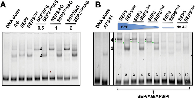
Figure 5.
Analysis of tetramer formation between SEP3 and SEP3Δtet and floral organ identity partners. (A) EMSA of SEP3, SEP3Δtet and AG with increasing protein concentrations (0.5, 1 and 2 μl of in vitro transcription-translation product) with a 103 bp fragment of the SEP3 promoter as per Figure 1. Lanes are as indicated and labeled. For single proteins, 2 μl of in vitro transcription-translation product was used. (B) EMSA of SEP3, SEP3Δtet with floral organ identity partners AP3, PI and AG with the same DNA as in (A). Lanes are as marked with decreasing concentrations of SEP3 and SEP3Δtet with equivolumes of SEP3 or SEP3Δtet and AG present in lanes 5 and 6 and no SEP3 or SEP3 Δtet present in lanes 7 and 8. In lanes 9 and 10, no AG was present but equivolumes of AP3, PI and SEP3 or SEP3Δtet were added. A total of 4 μl of in vitro transcription-translation product was used in all assays. 2 and 4 indicate number of protein molecules bound with green asterisks indicating SEP3 (or SEP Δtet)/AG/AP3/PI tetramer and black asterisks indicating SEP3/AG tetramers.

