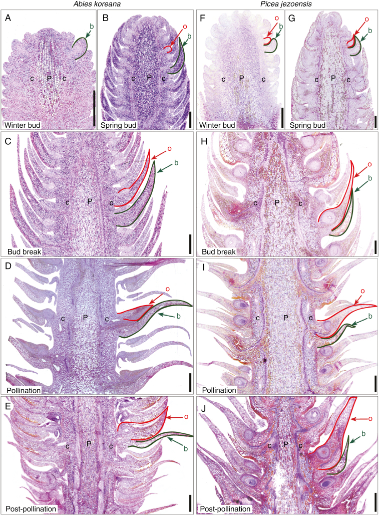
Fig. 2.
Seed cone anatomy and developmental in Abies koreana (A–E) and Picea jezoensis (F–J). (A) Abies cone before winter dormancy. Note that ovuliferous scale primordia have not formed. (B) Cone in early spring prior to bud break, showing initial differentiation of ovuliferous scale. (C) Cone in spring around bud break, when bract scales and ovuliferous scales are roughly equal in length. (D) Cone at pollination, showing spaces between bract scale/ovuliferous scale complexes and the large bract scale with small ovuliferous scale. (E) Cone following pollination, after ovuliferous scales have expanded to fill gaps between adjacent bract scale/ovuliferous scale complexes. (F) Picea cone before winter dormancy. Note that ovuliferous scale primordia are present. (G) Cone in early spring with more developed bract scale/ovuliferous scale complexes. (H) Cone during bud break. Note that ovuliferous scales have already elongated beyond the bract scales, whose cells are fully mature. (I) Cone at pollination showing enlarged ovuliferous scales and much smaller bract scales. (J) Cone after pollination. Note the expanded ovuliferous scale bases that have reoriented the bract scale/ovuliferous scale complexes into an imbricated arrangement. Abbreviations: b, bract scales (highlighted in green); c, cortical tissue; P, pith tissue; o, ovuliferous scales (highlighted in red). Scale bars (A–C, F–H) = 500 µm; (D, E, I, J) = 1000 µm.

