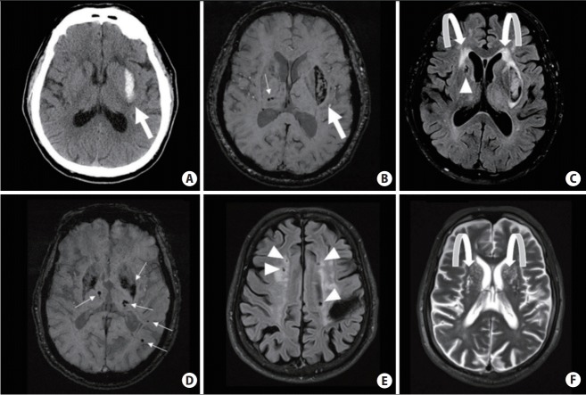
Figure 1.
Computed tomography and magnetic resonance imaging (MRI) scans of hypertensive small vessel disease and associated bleeding risks. (A–C) Hypertensive intracerebral hemorrhage (ICH) in the left basal ganglia (big arrows); ~1.6% to 2% annual recurrence risk. Several deep microbleeds (MBs) (small arrows), periventricular white matter hyperintensities (curved arrows) and a deep lacune (arrowhead) are also seen. (D–F) Mixed-location of ICH and MBs (small arrows), associated with ~5.1% annual risk of ICH recurrence. There are several lacunes (arrowheads) and MRI-visible enlarged perivascular spaces near the basal ganglia (curved arrows) in the same patient.

