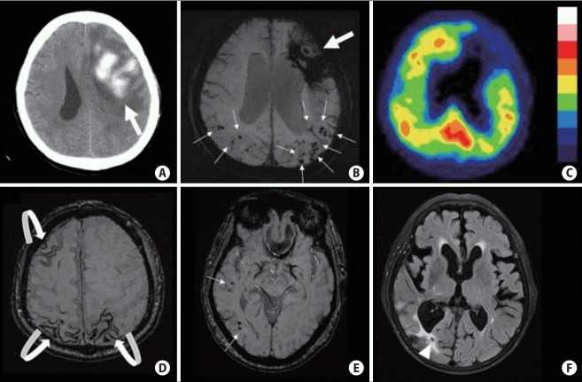
Figure 2.
Computed tomography and magnetic resonance imaging scans of cerebral amyloid angiopathy (CAA) related pathologies and associated bleeding risk. (A, B) Probable CAA (per Boston criteria) with lobar intracerebral hemorrhage (ICH) (big arrows) and multiple cortical microbleeds (MBs) (small arrows); ~10% annual ICH recurrence risk. (C) Increased tracer uptake detected on 11C-Pittsburgh Compound B positron emission tomography scans in the same patient as A and B note the posterior predominance typical of CAA. (D) Multifocal cortical superficial siderosis (curved arrows) in a probable CAA patient who had ICH (not shown on this cut); ~27% annual risk of recurrent ICH. (E) CAA presenting with only cortical MBs (small arrows); ~5% yearly risk of first-time symptomatic ICH. (F) Lobar lacune (arrowhead) in a probable CAA patient who had ICH (not shown on this cut).

