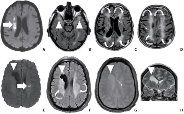
Figure 3.
Neuroimaging characteristics of two patients with hereditary small vessel diseases (SVD). Top row: A 64-year-old male patient without medical history presented with acute left hemiplegia. (A) Imaging revealed an acute right deep-seated infarct (arrow) as well as (B, C) widespread white matter hyperintensities (curved arrows, arrowheads) and (C, D) multiple lacunes (small arrows). (B) Fluid attenuated inversion recovery (FLAIR) also shows bilateral anterior temporal white matter hyperintensities (arrowheads). The patient was diagnosed with Cerebral Autosomal Dominant Arteriopathy with Subcortical Infarcts and Leukoencephalopathy (CADASIL) based on genetic testing. Bottom row: a 32-year-old male patient presented with acute dysarthria related to a small subcortical infarct (E, arrow). (F) Brain MRI showed otherwise diffuse white matter hyperintensities (curved arrows), (E–H) a right porencephalic lesion (arrowhead) and (G) microbleeds (small arrow). The patient was proven to have a COLIVA mutation.

