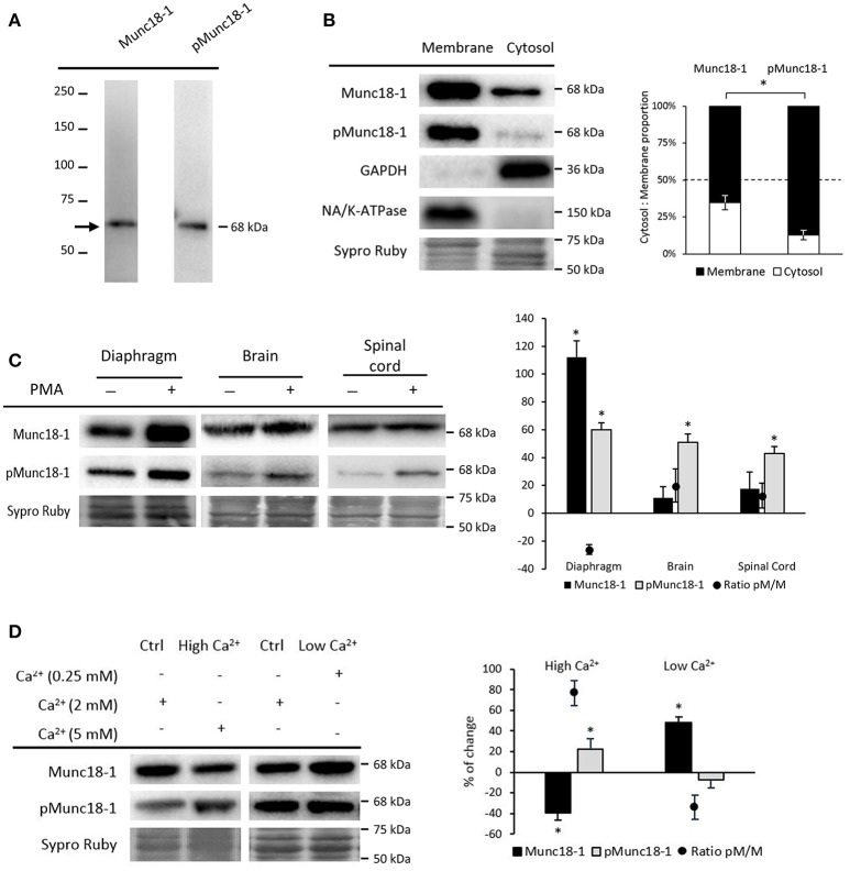
Figure 1.
Munc18-1 and pMunc18-1 in adult skeletal muscle under basal conditions. (A) Representative Western blot bands from diaphragm showing specificity for the antibodies Munc18-1 and pMunc18-1 (Ser-313). (B) Representative Western blot bands and quantification of membrane and cytosol distribution of Munc18-1 and pMunc18-1. Both are mainly located in the membrane but pMunc18-1 is barely detectable in the cytosolic fraction. GAPDH is exclusively in the cytosol fraction and the Na+/K+-ATPase is in the membrane. (C) Representative Western blot bands and quantification of Munc18-1 and pMunc18-1 after PMA treatment in diaphragm, brain and spinal cord. (D) Representative Western blot bands and quantification of Munc18-1 and pMunc18-1 after different calcium concentrations. High Ca2+ concentration increases pMunc18-1 levels while decreases Munc18-1. Data are mean percentage ± SEM, *p < 0.05 (n = 5).

