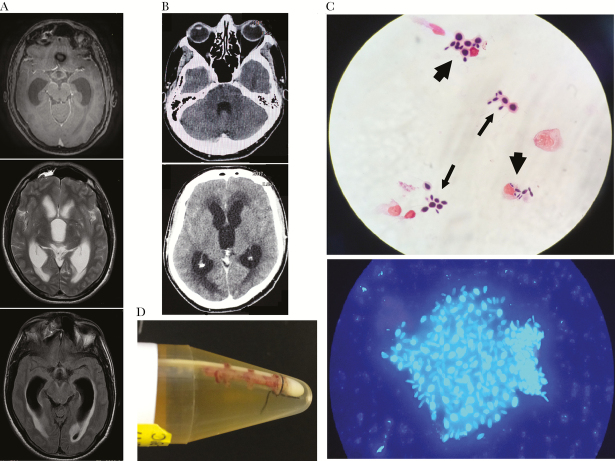
Figure 1.
Magnetic resonance imaging (MRI) and computed tomography (CT) scan of the brain and direct mycological exam of cerebrospinal fluid (CSF). A, Pretreatment (March 2016) brain T1-weighted MRI of patient 1 showing hydrocephalus and skull base meningeal contrast enhancement. B, Pretreatment (May 2017) CT scan of the brain of patient 2 showing communicating hydrocephalus and transependymal edema without anomalous intraparenchymal contrast enhancement or intraparenchymal lesions. C, Upper image: baseline CSF direct mycological exam (August 2017) of patient 2 showing yeast cells suggestive of Sporothrix sp. free (thin arrows) or engulfed by macrophages (thick arrows). Lower image: calcofluor white staining of patient 2’s CSF sample collected on August 2017, showing a yeast aggregate surrounded by extracellular matrix suggestive of a biofilm-like structure. D, fibrin sheath on the tip of the ventriculoperitoneal shunt removed (August 2017) from patient 2: culture of the material from the tip yielded Sporothrix sp., subsequently identified by molecular method as S. brasiliensis.

