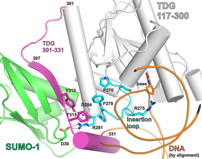
Figure 8.
Closeup view of the SUMO–SIM interface and the catalytic ‘insertion’ loop, using the same crystal structures and coloring scheme as described above for Figure 1B. Some of the residues that mediate SUMO–SIM binding, and residues of the catalytic ‘insertion’ loop are shown. Interactions between these residues could potentially link SUMO–SIM binding to impairment of productive nucleotide flipping. Because no structure is available for sumoylated TDG bound to DNA, the DNA shown (orange) was positioned as described for Figure 1B.

