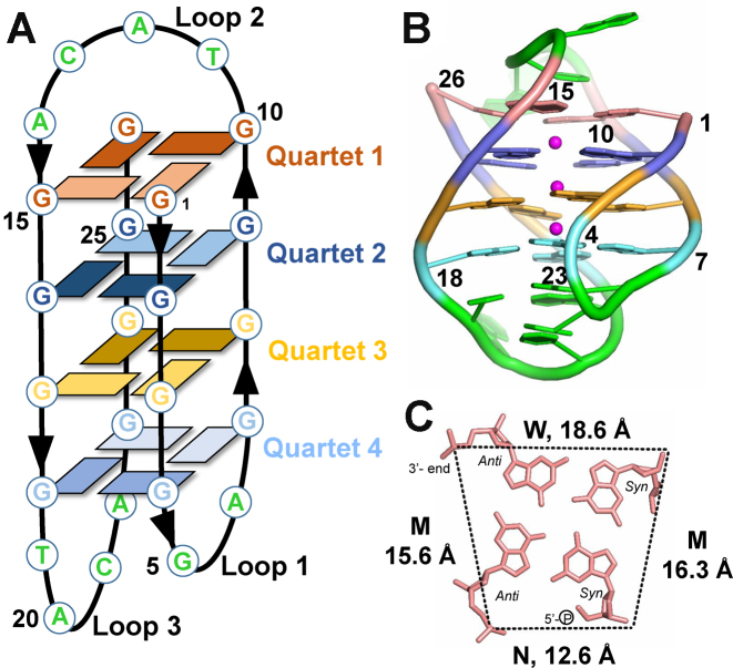
Figure 2.
Overall view of the D. discoideum G-quadruplex, 19wt. (A) Schematic representation of the G4. Dark and light rectangles indicate anti and syn conformations of guanine bases, respectively. Chain orientation is indicated by arrow heads. Each G-quartet is colored in a unique color throughout the figure. Loop nucleotides are colored in green. (B) Cartoon representation of the crystal structure with purines and pyrimidines shown as filled rings. Sugar rings are omitted for clarity. Same color coding is used as in panel A, with magenta spheres representing K+ ions. (C) Quartet 1 (G1⋅G10⋅G26⋅G15). N – narrow, M – medium, and W – wide grooves. Average groove width values are indicated. Phosphate-phosphate distances are shown as dashed lines. Nucleotides are depicted as sticks.

