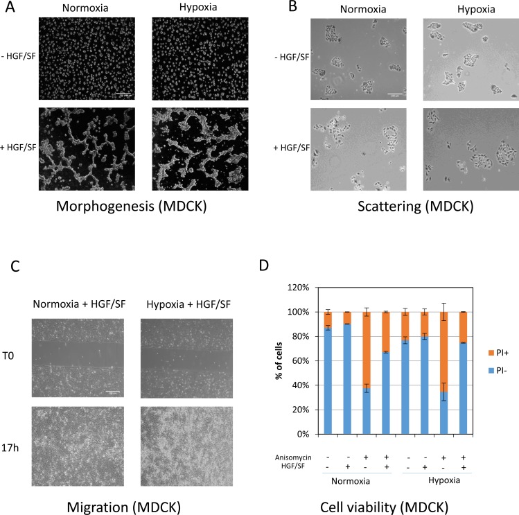
Figure 4. Effect of the MET phosphorylation decrease observed under hypoxia on biological responses induced by HGF/SF.
(A) MDCK cells were seeded on a layer of Matrigel™ for 24 h and then stimulated or not with 10 ng/mL HGF/SF, placed under normoxia or hypoxia for an additional 24 h, and photographed. The white scale bar corresponds to 200 μm. (B) MDCK cells were seeded and, after adhesion, serum-starved in the presence or absence of HGF/SF at 10 ng/mL. They were then placed under normoxic or hypoxic conditions for 24 h and fixed, stained with hematoxylin and eosin, and photographed. The white scale bar corresponds to 200 μm. (C) MDCK cells were seeded in an IBIDI® insert. The next day, they were treated with 10 µg/mL mitomycin-c to prevent proliferation. An hour later, the mitomycin-C was removed and the cells were left overnight under normoxic or hypoxic conditions. The cells were then photographed. White scale bar: 200 μm. (D) MDCK cells were seeded and serum-starved for 24 h after adhesion. The cells were then treated or not with 0.7 µM anisomycin in the presence or absence of 25 ng/mL HGF/SF. The next day, they were stained with propidium iodide (PI) for evaluation of cell viability.

