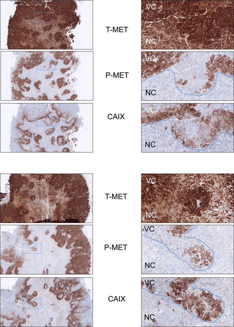
Figure 5. Decreased MET phosphorylation in hypoxic areas of GTL16 tumor xenografts.
GTL16 cells were xenografted subcutaneously into flanks of SCID mice. Tumors were analyzed by immunohistochemistry on 7-µm-thick sections with antibodies directed against the intracellular domain of MET, the phosphorylated form of MET, or the hypoxia marker CAIX. Two tumors are shown in this figure. Left panels: magnification factor ×4; right panels: magnification factor ×20 (applied to the boxed region). NC: Necrotic tumor cells. VC: Viable tumor cells.

