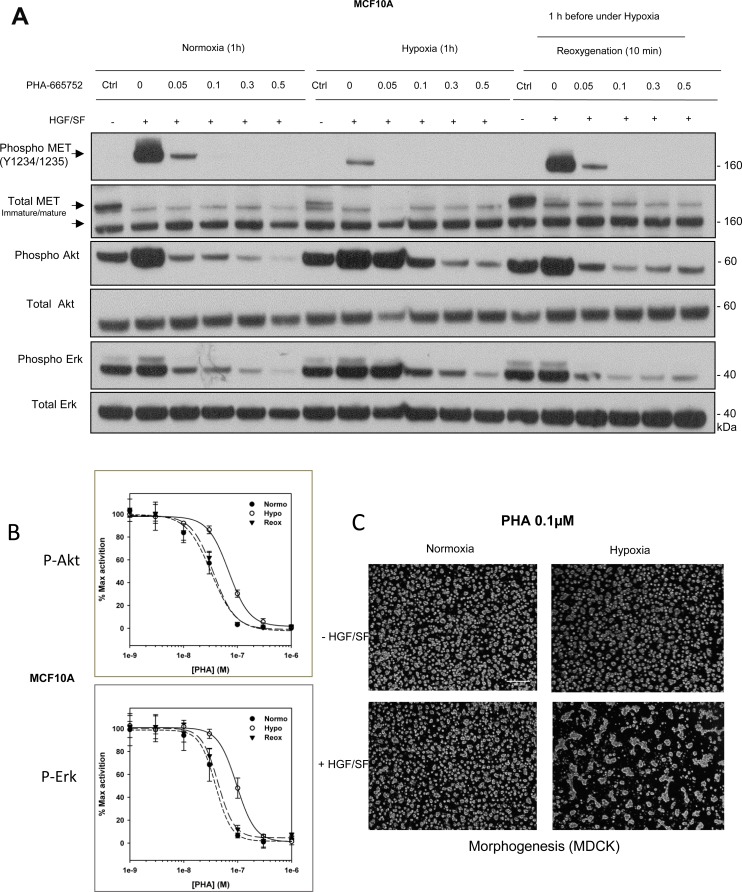
Figure 7. Responses to the tyrosine kinase inhibitor PHA-665752 under hypoxia and reoxygenation.
(A) MCF10A cells were treated with the MET tyrosine kinase inhibitor PHA-665752 at the indicated concentrations and then placed under normoxia or hypoxia for 1 h or under hypoxia for 1 h and then normoxia for 10 minutes (reoxygenation). The cells were then stimulated or not for 10 min with 10 ng/mL HGF/SF. The same amount of protein was resolved by 4–12% Bis-Tris SDS-PAGE and analyzed by western blotting with antibodies directed against phosphorylated residues of the MET kinase domain, the MET kinase domain, phosphorylated Akt, Akt, phosphorylated Erk, or Erk2. The positions of prestained molecular weight markers are indicated. Arrows indicate the positions of precursor and mature full-length MET. (B) MCF10A cells were placed under normoxic or hypoxic conditions for 1.5 h or hypoxia for 1.5 h and then normoxia for 10 minutes (reoxygenation) then treated for 10 minutes with HGF/SF 10 ng/mL and PHA-665752 at 10–3, 3.10–3, 10–2, 3.10–2, 0.1, 0.3 and 1 µM. A control without HGF/SF and/or PHA-665752 was included. Error bars represent standard deviations (n = 3; ± SD). (C) MDCK cells were seeded on a layer of Matrigel™ for 24 h and then stimulated or not with 10 ng/mL HGF/SF, with or without 0.1 µM PHA-665752. They were then placed under normoxia or hypoxia for an additional 24 h. Finally they were photographed. White scale bar: 200 μm.

