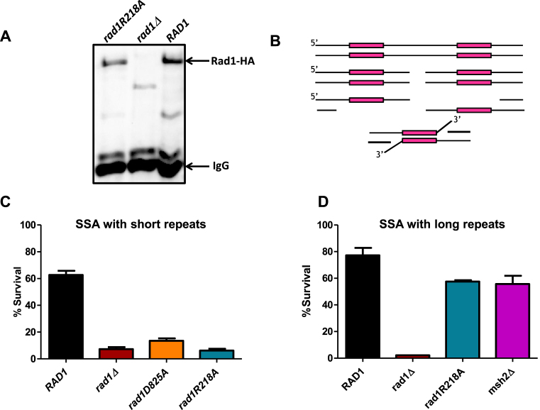
Figure 8.
rad1R218A function in SSA. (A) rad1R218A-3HA was integrated into the chromosome, replacing endogenous RAD1. To determine expression levels, α-HA antibody was used to immunoprecipitate HA-tagged protein from cell lysates grown to mid-log phase for rad1R218A-3HA, RAD1–3HA, and rad1Δ w. A western blot of eluates was performed using α−HA antibody. rad1Δ serves as a negative control. An arrow indicates the Rad1–3HA or rad1R218A-3HA band. (B) Cartoon of SSA in the EAY1141 (short ∼250 bp ura3 repeats) and YMV80 (long ∼ 1.3 kb leu2 repeats) strains used to assess 3′ NHTR.(C) Msh2–Msh3-dependent SSA assays with short repeats were performed. D. Msh2–Msh3-independent SSA assays with long repeats were performed. Percent survival (induced/uninduced) was calculated by determining the viability of cells after a 5 hour induction of HO expression. Data represents the mean ± SEM of at least six independent experiments with at least two independent isolates. The following strains were used: RAD1 (black), rad1Δ (dark red), rad1D825A (orange), rad1R218A (teal) and msh2Δ (magenta).

