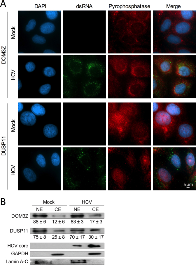
Figure 7.
DOM3Z and DUSP11 partially localize to the cytoplasm in Huh-7.5 cells. (A) Huh-7.5 cells were plated onto 8-well chamber slides and infected with JFH-1T (MOI = 0.1). After 3 days, cells were fixed and stained for dsRNA, DOM3Z (top panel) or DUSP11 (bottom panel) and DAPI. (B) Huh-7.5 cells were infected with JFH-1T (MOI = 0.1) and harvested 3 days post-infection. Following subcellular fractionation, cellular localization of the pyrophosphatases was determined by western blot with antibodies against DOM3Z and DUSP11. GAPDH and Lamin A–C were used as cytoplasmic and nuclear markers, respectively. HCV core was used to confirm HCV infection. Percent expression ± standard deviation relative to total expression is indicated. All data are representative of three independent experiments.

