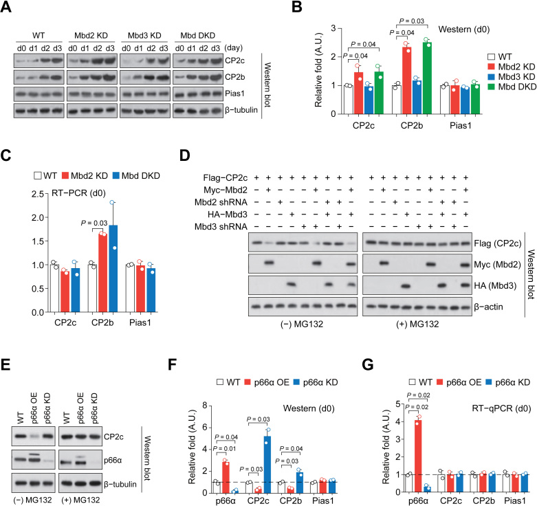
Figure 5.
p66α- and Mbd2-dependent degradation of CP2c and CP2b. (A) Immunoblot analysis of the CBP proteins expression in differentiating Mbd2 KD, Mbd3 KD and Mbd DKD MEL cells in vitro. (B) Quantification of the CBP proteins expression in uninduced cells in (A). n = 2. (C) RT-PCR analysis of the expression of CBP mRNAs in the uninduced Mbd2 KD and DKD cells. Values of uninduced cells shown in Supplementary Figure S5C are highlighted. n = 2. (D) Immunoblot analysis of CP2c protein expression in 293T cells transiently transfected of the Mbd2, Mbd3, shMbd2 and shMbd3 expression vectors, in combination, in the presence or absence of proteasome inhibitor MG132. (E) Immunoblot analysis of the CP2c expression in undifferentiated p66α OE and KD MEL cells in the presence or absence of MG132. (F and G) Immunoblot (F) and RT-qPCR (G) analyses of the CP2c, CP2b, and Pias1 expression in uninduced p66α OE and KD MEL cells. Values in uninduced cells shown in Supplementary Figures S5F and S5H are highlighted. n = 2.

