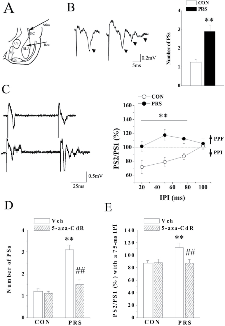
Figure 6.
DNA methyltransferase 1 (DNMT1)-mediated GABAergic deficit leads to the potentiation of cortical-basolateral amygdala (BLA) transmission in postnatal stress (PRS) mice. CON and PRS represent control and PRS mice, respectively. (A) Schematic diagram illustrating the placement of stimulating electrode (Stim) and recording electrode (Rec) at cotical-BLA pathway. (B) Left: Basal evoked synaptic response in the BLA of control and PRS mice. PS wave is marked with arrows. Right: The comparison of population spike (PS) number between control and PRS mice. (C) Left: The original results of paired-pulse stimulation with the interval at 50 ms. Right: The comparison of PS2/PS1 with the stimulus interval from 20 to 100 ms between control and PRS mice. Dotted line indicates 100%. ↑ and ↓ represent paired-pulse facilitation (PPF) and paired-pulse inhibition (PPI), respectively. (D and E) Effects of 5-aza-CdR on the number of PSs or the PPI induction in control and PRS mice. All bars represent means±SEM; 2-tailed Student’s t test for B and 2-way ANOVA followed by PLSD posthoc test for C, D, and E; **P<.01 between the data of control (n=10) and PRS mice (n=10) or between the data of vehicle-treated control (n=12) and vehicle-treated PRS mice (n=10). ##P<.01 between the data of vehicle-treated PRS mice (n=10) and 5-aza-CdR-treated PRS mice (n=10).

