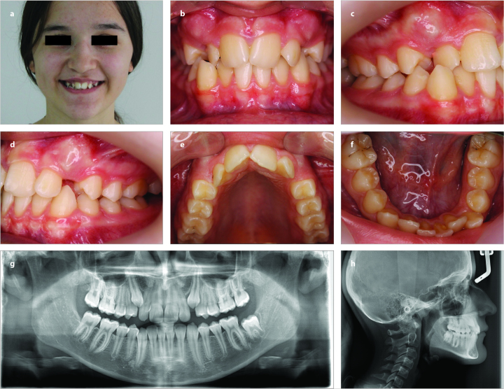
Figure 1. a–h.
Pretreatment-intraoral and extraoral photographs, lateral cephalometry, and panoramic radiograph; pretreatment photograpghs of the patients (a); intraoral frontal image (b); intraoral right image (c); intraoral left image (d); intraoral photographs of the patients (e); intraoral photographs of the patients (f); pretreatment panoramic film (g); pretreatment lateral cephalometric film (h)

