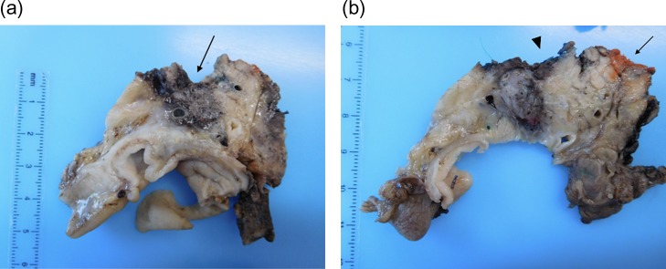
Figure 4:
(a and b) Pancreaticoduodenectomy specimen sliced in an axial plane, according to the Leeds Pathology Protocol, providing good visualization of the tumour and its relationship to key anatomical structures and the anterior, posterior and superior mesenteric vein groove surfaces. (a) Demonstrates an area of haemorrhage/necrosis due to IRE needles extending to the SMV bed (arrow) and (b) shows the SMV (arrrowhead) and the SMA groove (arrow).

