
Fig. 1. Organization and glycosylation of the LASV trimer.
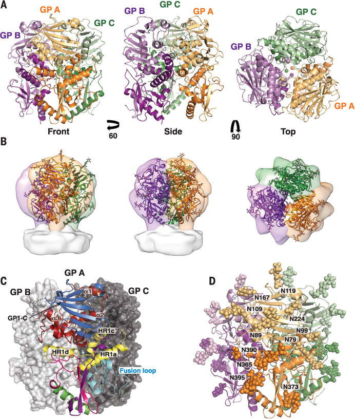
(A) Cartoon representation of the trimer from the front, side, and top. The GP1 subunit of each monomer is in a light shade and the GP2 subunit in a dark shade. In the top view, spheres indicate positions of the C terminus of GP1 and the N terminus of GP2 at the trimeric interface. (B) The crystal structure of the LASV GP trimer (cartoon) docked into the tomographic reconstruction of the LASV GPC spike from fixed virions (surface, EMD-3290), in the same orientations as shown in (A). (C) GP monomer A is shown in cartoon representation and is colored by domain as in fig. S3. Structural elements involved in trimerization are indicated. GP monomers B and C are shown as surfaces. (D) The glycans visible in the crystal structure are illustrated as atomic spheres, with those glycans attached to GP1 illustrated in light shades and those attached to GP2 illustrated in dark shades.