
Fig. 4. Structural definition of the 37.7H epitope.
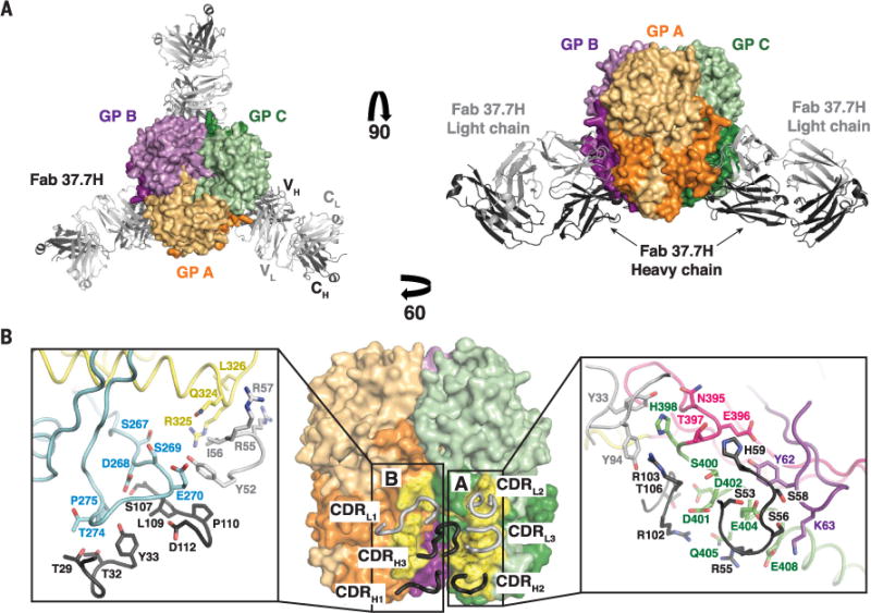
(A) The GP trimer bound to three 37.7H Fabs from the top (left) and side (right). Cartoon representations of the 37.7H Fab heavy and light chains are shown in dark and light gray, respectively. Each Fab binds to two GP monomers, shown as surface representations, near the base of the trimer. (B) Monomers A and C are shown as surface representations, with the heavy- and light-chain CDRs shown as dark and light gray tubes, respectively. The footprint of the antibody is colored yellow, with the binding site indicated. Side-chain interactions at the GP-37.7H interface are magnified in the inset boxes. GP elements are colored as indicated in fig. S3. Note that only selected residues are shown for clarity.