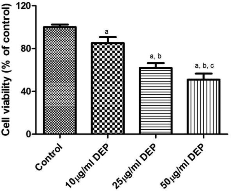
Fig. 1.
Cytotoxicity of DEPs on HBMVEC cells. Cells were treated with various concentrations of DEPS (10 µg/ml, 25 µg/ml, and 50 µg/ml). After 24 h of treatment, the cell viability was assessed by MTS assay. Values represent mean±SD. All experiments were performed in triplicates, and the values reported are mean ± SD. (a: different from control group, b: different from 10 µg/ml [DEP] group, and c: different from 25 µg/ml [DEP] group, p<0.05).

