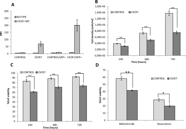
Fig 4. Effect of CXCR7 receptor overexpression on U2932 DLBCL cells.
(A) Quantification of CXCR7 expression by flow cytometry, in the total population of transfected U2932-CXCR7 and U2932-control cells or after measurements restricted only to GFP+ cells. Values are expressed as mean fluorescence intensity (MFI) (B) Proliferation rate as measured by cell density (number of cells/ml) over time in GFP+ U2932-CXCR7 or U2932-control cells (C) Percentage of cell viability, measured by Trypan Blue staining, over time for GFP+ U2932-CXCR7 or U2932-control cells (D) Antitumor effect, expressed as percentage of cell viability respect to control cells, after 48h exposure of GFP+ U2932-CXCR7, or U2932-control cells, to 10μM mafosfamide or 1μM doxorubicin. ** p<0,01; * p<0,05.

