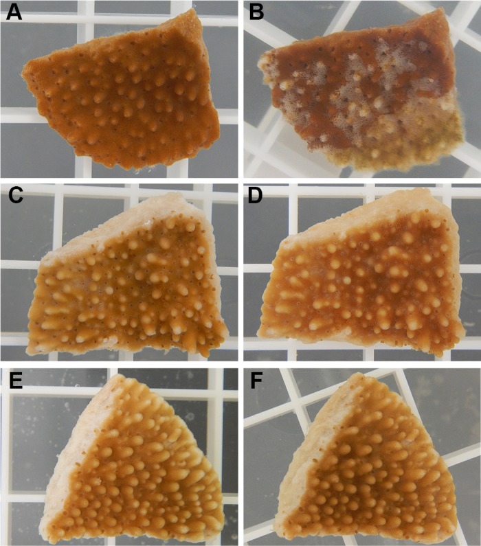
Fig 6. Representative photographs of M. capitata fragments used in infection experiments.

(A) A coral fragment before exposure to OCN008, and (B) the same coral fragment 24 h post-exposure to OCN008 exhibiting extensive tissue loss/lysis. (C) A fragment before exposure to the ΔompU strain of OCN008, and (D) the same fragment 240 h post-exposure to the ΔompU strain. (E) A fragment before exposure to the control bacterium HMSC5, and (F) the same fragment 240 h post-exposure to HMSC5. The white square grating measures 1 x 1 cm.
